Glaucoma: Diode Laser Treatment
________________________________________________________________________
KEY POINTS
- Diode laser treatment uses a laser to destroy part of the eye that makes fluid in people with glaucoma. This procedure reduces the fluid the eye produces, and thus lowers eye pressure. In some cases, it also relieves eye pain. Controlling the pressure may reduce the risk of permanent blindness and help you keep your vision.
- Ask your provider how long it will take to recover and how to take care of yourself at home.
- Make sure you know what symptoms or problems you should watch for and what to do if you have them.
________________________________________________________________________
What is diode laser treatment?
This procedure uses a laser to reduce the amount of fluid an eye can produce. It destroys part of the eye that makes fluid. This treatment is also called a cyclodestructive procedure.
Glaucoma is an eye disease that damages the nerve that carries visual messages to the brain (optic nerve). This is usually caused by high pressure inside the eye. Damage to the optic nerve can cause a permanent loss of vision. Glaucoma needs to be diagnosed and treated early to prevent blindness.
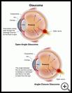
Normally, the fluid in the front of the eye (the aqueous humor) is constantly flowing from where it is formed (the ciliary body) to the front of the eye. This fluid nourishes your eye and helps to keep its shape. The area between the iris (colored part of the eye) and the cornea (the clear outer layer on the front of the eye) is called the angle. Fluid drains out through the angle, into drainage channels, and is then reabsorbed by the body. When fluid flows out too slowly, eye pressure builds up.
There are 2 main types of glaucoma.
In open-angle glaucoma, fluid drains slowly, causing the pressure in the eye to increase. This happens even though the drainage channels for the fluid are open. One type of open-angle glaucoma is caused by injury to the eye. In some cases of open angle glaucoma, it is not known what causes the fluid to drain out too slowly.
In angle-closure glaucoma, the angle between the iris and the cornea is blocked or narrowed. When this happens, fluid is not able to drain from the eye. This can cause a pressure buildup. This can happen if the pupil is dilated too much, causing the iris to "bunch up," or if the lens” crowds” the iris and causes it to bend forward and close the angle. When this type of glaucoma happens suddenly, it is called acute angle-closure glaucoma and is a medical emergency.
When is it used?
This procedure reduces the fluid the eye produces, and thus lowers eye pressure. In some cases, it also relieves eye pain. Controlling the pressure may reduce the risk of permanent blindness and help you keep your vision.
This procedure is most often done when other treatments have not worked, or would not be safe. In rare cases, laser is the first treatment for people who have severe glaucoma.
Ask your healthcare provider about your choices for treatment and the risks.
How do I prepare for this procedure?
- Plan for your care and a ride home after the procedure.
- You may or may not need to take your regular medicines the day of the procedure. Tell your healthcare provider about all medicines and supplements that you take. Some products may increase your risk of side effects. Ask your healthcare provider if you need to avoid taking any medicine or supplements before the procedure.
- Your healthcare provider will tell you when to stop eating and drinking before the procedure. This helps to keep you from vomiting during the procedure.
- Do not wear eye makeup on the day of the surgery.
- Follow any other instructions your healthcare provider gives you.
- Ask any questions you have before the procedure. You should understand what your healthcare provider is going to do. You have the right to make decisions about your healthcare and to give permission for any tests or procedures.
What happens during the procedure?
You will be given local anesthesia to keep you from feeling pain during the procedure. If you are having local anesthesia, you will be given medicine to help you relax, but you may be awake during the procedure. Then the provider will numb your eye by injecting an anesthetic around your eye.
Your provider will use a laser on the outside surface of the eye to destroy part of the eye that makes the fluid. The procedure will take 5 to 15 minutes once your eye is numb. If you are awake during the procedure, and a laser is used, you may hear a popping sound when the laser is on.
In some cases, laser treatment is done surgically from the inside of the eye. This is more common if you are having other surgery on that eye at the same time.
What happens after the procedure?
After the procedure the provider will put in eye drops or ointment and place a patch on your eye. You will be given a prescription for eye drops and sometimes for pain medicine. You will also need to schedule a follow-up appointment. Your vision may be blurry and you may have some pain while your eyes heal.
Ask your healthcare provider:
- How long it will take to recover
- If there are activities you should avoid
- How to take care of yourself at home and when you can return to your normal activities
- What symptoms or problems you should watch for and what to do if you have them
Make sure you know when you should come back for a checkup. Keep all appointments for provider visits or tests.
What are the risks of this procedure?
Every procedure or treatment has risks. Some possible risks of this procedure include:
- It may produce a cataract, which is a cloudy lens in the eye. This can cause blurry vision. Often cataracts can be treated with surgery.
- It may cause swelling of the retina. Often this can be treated with medicines.
- It may make eye pressure too low. This is a very difficult problem that can lead to vision loss.
Ask your healthcare provider how these risks apply to you. Be sure to discuss any other questions or concerns that you may have.

