Glaucoma: Surgery to Lower Eye Pressure
________________________________________________________________________
KEY POINTS
- The two main types of glaucoma surgery are trabeculectomy and tube shunt surgery. Both create small passageways from the inside to the outside of your eye in order to better drain the fluid from your eye.
- Glaucoma surgery can lower the pressure in your eye and may help prevent more damage to the optic nerve and further loss of vision.
- Ask your provider how long it will take to recover and how to take care of yourself at home.
- Make sure you know what symptoms or problems you should watch for and what to do if you have them.
________________________________________________________________________
What is glaucoma surgery?
Glaucoma is an eye disease that damages the nerve that carries visual messages to the brain (optic nerve). This is usually caused by high pressure inside the eye. Damage to the optic nerve can cause a permanent loss of vision. Glaucoma needs to be diagnosed and treated early to prevent blindness.
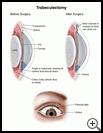
Normally, the fluid in the front of the eye (the aqueous humor) is constantly flowing from where it is formed (the ciliary body) to the front of the eye. This fluid nourishes your eye and helps to keep its shape. The area between the iris (colored part of the eye) and the cornea (the clear outer layer on the front of the eye) is called the angle. Fluid drains out through the angle, into drainage channels, and is then reabsorbed by the body. When fluid flows out too slowly, eye pressure builds up.
The two main types of glaucoma surgery are trabeculectomy and tube shunt surgery. Both create small passageways from the inside to the outside of your eye in order to better drain the fluid from your eye. In trabeculectomy, the passageway is made out of the white part of the eye. In tube shunt surgery, a tiny tube is placed in the eye. Both can lower the pressure in your eye and may help prevent more damage to the optic nerve and loss of vision.
When is it used?
Trabeculectomy (also called filtering surgery) or tube shunt surgery may be used when:
- Medicines do not lower your eye pressure enough.
- You are having harmful side effects from medicines you are taking for the pressure in your eyes or cannot successfully use the medicines your provider prescribes.
- Laser surgery to lower the eye pressure has not worked or is not possible.
Ask your healthcare provider about your choices for treatment and the risks.
How do I prepare for this procedure?
- Plan for your care and a ride home after the procedure.
- You may or may not need to take your regular medicines the day of the procedure. Tell your healthcare provider about all medicines and supplements that you take. Some products may increase your risk of side effects. Ask your healthcare provider if you need to avoid taking any medicine or supplements before the procedure.
- Your healthcare provider will tell you when to stop eating and drinking before the procedure. This helps to keep you from vomiting during the procedure.
- Do not wear eye makeup on the day of the surgery.
- Follow any other instructions your healthcare provider gives you.
- Ask any questions you have before the procedure. You should understand what your healthcare provider is going to do. You have the right to make decisions about your healthcare and to give permission for any tests or procedures.
What happens during the procedure?
The procedure is done in an operating room.
You will be given a sedative to help you relax. Your eye will be numbed with eye drops or a shot of a local anesthetic around the eye so you will not feel pain during the surgery. Some people need general anesthesia. General anesthesia relaxes your muscles and you will be asleep.
After you have been given the anesthetic, your provider will make a small drainage channel so that fluid can flow from inside the eye to outside the eye. The fluid flows through the new opening, under the clear covering over the white part of your eye, and then collects in a tiny pouch, called a bleb, underneath the eyelid. The fluid is then slowly absorbed into the bloodstream. Your provider may put some medicine on the eye to keep the new channel from scarring closed.
What happens after the procedure?
You will need to be examined the next day. Your provider may want to check your eyes once or twice a week for 4 to 6 weeks after your surgery.
You will use various eye medicines after surgery to help healing and to reduce the risk of infection.
Your vision may be blurred for several weeks after surgery. You may need medicine after the surgery to help maintain normal eye pressure. Once your eye heals, you may need to get a new eyeglass prescription.
In general, you will not be able to wear contact lenses after trabeculectomy. If you are a contact lens wearer, talk to your eye care provider about your options.
Ask your healthcare provider:
- How long it will take to recover
- If there are activities you should avoid
- How to take care of yourself at home and when you can return to your normal activities
- What symptoms or problems you should watch for and what to do if you have them
Make sure you know when you should come back for a checkup. Keep all appointments for provider visits or tests.
What are the risks of this procedure?
Every procedure or treatment has risks. Some possible risks of this procedure include:
- Developing a cataract, which is a cloudy lens in the eye.
- Scarring around the new drainage site, causing the pressure to increase and sometimes requiring a second surgery.
- Bleeding inside or around your eye. A little blood in your eye is common and usually does not need treatment. Severe bleeding is rare but can cause permanent vision loss.
- Getting an infection. This can happen soon or long after the surgery. Call your provider right away or go to the emergency room if you have decreased vision, pain, redness, or drainage in the eye that was treated.
- Too much drainage and low eye pressure. This sometimes goes away on its own. Sometimes further surgery is needed.
Ask your healthcare provider how these risks apply to you. Be sure to discuss any other questions or concerns that you may have.

